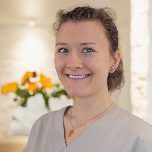
Dr. Josephine Phillips
- Oral surgeon
- Specialist for ceramic implants
- Specialist in biological dentistry
- Extensive experience in implantology
- Focus on minimally invasive surgical techniques
- Part of our team since 2019
Dr. Josephine Phillips (neé Dr. Josephine Tietje) joined our practice in 2019. She is oral surgeon and specialist in biological dentistry and ceramic implantology, performing all types of dental surgery.
Experienced, confident and enthusiastic for her field, she removes inflamed teeth and jaw inflammations with minimally invasive techniques – protecting the jaw bone to maintain a healthy basis for both immediate and late implants. She performs bone augmentations such as internal and external sinus lifts, three-dimensional horizontal and vertical bone augmentation techniques using the patient’s own bone. During difficult tooth extractions, she carefully removes teeth while gently handling the tissues, so that impacted wisdom teeth or cysts lying close to the nerve can be removed without complications.
Career: Dr. Phillips studied dentistry at the University of Muenster in Germany from 2007 to 2013. She passed the state examination as the best of her semester. She completed several internships at the University Clinic Basel.
During her doctoral studies, she focused on minimally invasive vascular surgical techniques. She then specialized in oral surgery, practicing in three renowned surgical clinics and successfully completed the oral surgical examination in 2018.
Since then, Dr. Phillips has been practicing purely biological dentistry, performing all types of surgical treatments – from wisdom tooth extractions to complex implant-supported complete restorations, cystectomies, foreign body removals, treatments of chronic inflammations and infections of bone and soft tissue, incorporating various methods of biological dentistry – e.g. neural therapy, ART testing by Dr. Klinghardt, A-PRF and ozone.
Of course, the periodontal ligament is removed thoroughly.
She is a certified specialist in Biological Dentistry and Ceramic Implantology and regularly holds speeches for Swiss and international expert audiences, presenting her skills during live surgeries and articles in professional journals.
With passion for surgery and a calm treatment atmosphere, Dr. Phillips is always there for our patients, she is careful and takes time for your individual situation. We are happy to advise you regarding possible dental or surgical treatment options in your case.
We would like you, as our patient, to begin your healing journey in complete peace and relaxation.
We are here to assist you
Our dental practice is located directly at the Winterthur train station, above the Santa Lucia restaurant. If you arrive by train and exit the main train station, the building is on the left, a minute's walk away. You can park your car in the Stadtgarten (Coop/Manor) parking garage, which is only a few meters from the practice. Patients arriving from Zurich Airport can easily reach us by train in about 13 minutes.
Dentist Winterthur
8400 Winterthur
Switzerland
Office hours
9 – 17 h
By appointment only